TISSUE
Tissue is a cellular organizational level intermediate between cells and a complete organism. A tissue is an ensemble of similar cells from the same origin that together carry out a specific function. Organs are then formed by the functional grouping together of multiple tissues.
The study of internal structure of plants /animal called anatomy includes histology; that is organization and structure of tissue. Different type of tissue have different physical properties. Tissues may be hard (bone), soft (muscle) of liquid (blood). In this chapter we will learn different type of tissue present in plant and animal and their structural details.
- In unicellular organisms a single cell performs all the vital activities for example, digestion, respiration, excretion etc.
- In case of Multicellular organisms specialized functions are performed by a different groups of cells. As blood flows for transportation of O2, CO2, food hormones & waste material, muscle cells are involved in movement etc.
- In plants vascular tissue conduct food & water from one plant to another par to the plant Thus Multicellular organisms possess well-developed division of provide highest possible efficiency or particular function.
- A tissue is defined as a group of cells with similar structure, organized to do a common function.
- Term tissue was coined by Bichat.
- As plants are fixed or stationary, most of their tissues are of supportive type. Animals move around in search of food, mate & shelter so they consume more energy as compared to plants.
- Plants have some localized regions with special tissue but there are no such distinct regions in animals. Growth in animals remains uniform. Branch of biology deals with the study of tissue is called Histology.
DIFFERENCES BETWEEN THE TISSUES OF PLANTS AND ANIMALS:
| S.No. | Plant tissues | Animal tissues |
| 1. | Most of the plants remain fixed at one place (sedentary) and need less energy. Therefore the tissues are thick – walled and dead to provide mechanical support. E.g., Sclerenchyma. | Animals show active locomotion and hence need more energy. Therefore the tissues are made up of living cells. E.g. – Nervous tissue. |
| 2. | Growth in plants is indefinite and limited to certain regions. Certain tissues (meristematic) in root and shoot apex divide throughout life and add new cells. These cells differentiate and stop dividing to form permanent tissues. | Growth in animals is definite and not limited. In other words, they do not possess dividing and non-dividing tissues. The animal organs grow more or less uniform. |
| 3. | Plant tissues are not much complicated. | Animal tissues are much more complicated. |
A multicellular organism (plants and animals) develops from single ‘initial cell’ which undergoes repeated cell division. The large number of cells so formed undergoes cellular differentiation (a process of qualitative changes in the cells to perform different functions in the living organisms). Cell division and differentiation leads to the development of specific organs, each, consisting of specific groups of cells to perform specific functions in the body. With the increase in complexity of organisms, the level of organization also becomes complex.

Plant Tissues
- Meristematic tissue
- Permanent tissue
This differentiation is based on the ability of the mature cells of the tissue to divide and produce new cells. Meristematic tissue cells are capable of dividing, while permanent tissue cells are not.
Meristematic tissues
Meristematic tissues are seen in plants. They are primarily made up of rapidly dividing cells. They are the growing tissues of the plant.
Permanent tissues
Permanent tissues arise from the meristematic tissue and have structural and functional properties. Permanent tissue can be made up of either living or dead cells. They are specialised to perform a specific function, e.g. parenchyma, collenchyma, xylem, phloem, etc. Permanent tissues are of two types simple and complex permanent tissue.
Differentiation
Differentiation is the process by which the meristematic tissues develop into different types of permanent tissues based on the location and requirements of the plant.
Apical meristem
Apical meristem is present on the apex of the plant shoot and root. They are rapidly growing tissues and aid in increasing the height of the plant.
Lateral meristem
Lateral meristem is present on the lateral walls of the stem. They help in the horizontal growth of the plant and increase the stem girth.
Intercalary meristem
Intercalary meristem can be found between the nodes of the stem and the base of the leaf. They help in branching.
PERMANENT TISSUES
- The permanent tissues are composed of those cells which have lost their capability to divide. They have definite shape, size and thickness. The permanent tissue may be dead or living.
- The division & differentiation of the cells of Meristematic tissues give rise to permanent tissues. In cell differentiation, developing tissue and organs change from simple to more complex forms to become specialized for specific functions. The cells of permanent tissue loose the capacity to divide and attain a permanent shape, size and function.
CHARACTERISTICS OF PERMANENT TISSUES:
- The cells have lost their power of division.
- The cells possess definite shape, size and function.
- They may be living or dead.
- The living permanent cells are large, thin walled with a vacuolated cytoplasm.
- Dead permanent cells are thick walled without cytoplasm.
TYPES OF PERMANENT TISSUES:
The permanent tissues are classified on the basis of their composition into two types.
- simple permanent tissue
- Complex permanent tissue
- Simple permanent tissues: on the basis of structure of constituent cell, Simple tissues are of three types Parenchyma, Collenchyma and sclerenchyma.
Parenchyma
Parenchyma (Gk. para = beside; en-chein = to pour; i.e., some semi liquid substance poured beside other solid tissues; Grew 1682) is the most basic type of differentiated tissue from which other types have evolved.
Parenchyma

Characteristics
- The cells are living and may be rounded, oval, rectangular, star shaped but usually polygonal. Number of sides in a polygonal cell is usually 14 but in Elodea, it may be 17.
- Cells are loosely arranged with many intercellular spaces, either schizogenous or lysigenous (exception epidermis/epiblema, endodermis, pericycle and pith rays, where cells are compactly arranged).
- The cell wall is thin and is made up of cellulose, hemicellulose and pectin. Though the cell wall of xylem parenchyma is thick, that of epidermal cells is cutinised and that of endodermal cells is suberised.
- Cytoplasm is dense, granular with a central, large prominent nucleus.
- Vacuoles are many but small.
- Cytoplasm has all the cell organelles but instead of chloroplasts, there are leucoplasts.
Modifications of parenchyma
To perform functions other than normal (storage of food), parenchyma gets modified into following types:
- Chlorenchyma: Parenchyma cells having chloroplast; found in mesophyll of leaves; meant for photosynthesis.
- Aerenchyma: Parenchyma with air filled spaces; found in hydrophytes, meant for buoyancy.
Origin
- Parenchyma originates from the cells of meristem by loosening their divisional capacity.
functions
- Storage of food is the main function.
- Storage of water especially in xerophytes.
- Parenchyma associated with the vascular tissues (xylem and phloem) plays an important role in the conduction of sap and transportation of food.
COLLENCHYMA
- Collenchyma [Gk. Colla: glue, referring to its characteristic shining wall; Schleiden, (1839)] is a specialized supporting tissue which has living cells and possesses considerable tensile strength.
- It is usually found in stems beneath the epidermis as a complete cylinder or in the form of longitudinal strips.
STRUCTURE
- Cells of collenchyma are elongated, tubular and are arranged along the long axis of the stem but in T.S. they appear as circular, oval or polyhedral.
- The peculiarity of collenchyma cells is the unevenly thickened cell wall i.e., thickenings are restricted to the corners of the cells.
- The thickening materials are mostly pectin in addition to cellulose and hemicellulose.
- The cells are living and the protoplast is highly vacuolated. Therefore, cytoplasm and nucleus become peripheral.
- Cytoplasm has all the cell organelles including chloroplasts.
DIFFERENT TYPES OF COLLENCHYMA
- On the basis of mode of wall thickening, collenchyma may be classified into the following three types:
Lamellar : In this type, the thickenings are more heavily deposited on the tangential than on the radial cell walls. This type of collenchyma occurs in the stem of Raphanus, Helianthus, Rheum, etc.
Angular : In this type, the thickenings are primarily deposited at the corners or angles of the cells. Angular collenchyma, the most common type of collenchyma is found in the stems of Datura, Lycopersicum, Cucurbita, Solanum, Ficus, Vitis, Morus, Polygonum, etc.
Lacunar : In this type, the thickenings are primarily deposited around the intercellular spaces, e.g., in aerial roots of Monstera and petioles of Malva, Asclepias, etc.
Lamellate collenchyma Angular collenchyma Lacunate collenchyma

FUNCTIONS
❒ Collenchyma performs the following functions:
It is simple living mechanical tissue which provides mechanical support.
Being flexible in nature, it provides tensile strength to the plant body.
As the cells of collenchyma are living and often contain chloroplasts they also take part in photosynthesis.
Differences between parenchyma and collenchymas:
| S.No. | Parenchyma | Collenchyma |
| 1. | It consists of thin-walled living cells. | It consists of cells with localized thickenings. |
| 2. | It is distributed in all plant parts. | It is found in aerial parts and restricted to outer layers. |
| 3. | The cells of parenchyma assimilate, store food and waste products. | Collenchyma forms the mechanical tissue in young parts of the plants. |
SCLERENCHYMA
- Sclerenchyma (Gk. Scleros: hard; Mettenius 1805) is also a simple permanent tissue composed of thick walled, dead often lignified and hard cells. There is considerable variation in shape, size, origin and development of the cells.
- The mature sclerenchyma cells differ from parenchyma and collenchyma cells in the presence of lignified secondary walls and the absence of living protoplasts.
Different types of Sclerenchyma
- Fibres and sclereids are the two common categories of sclerenchyma.
Fibres
- They are specialized sclerenchymatous cells that occur in different parts of the plant body in bands or in the form of uninterrupted hollow cylinder.
They have great tensile strength, flexibility and elasticity which enable plant organs to withstand a variety of strains and tensions caused by the action of gravity, wind, etc.
Structure
- Fibres are long, narrow, thick and lignified sclerenchyma cells, usually with pointed or blunt ends. In transverse section, they appear rounded or polygonal in outline with a well defined lumen.
- Pits are few and simple, except fibre tracheids.
- Fibres are always found in sheets.
- Fibres are perhaps the longest cells among plant kingdom.
Sclereids
- Sclereids (Tschierch, 1885) are widely distributed and may occur almost anywhere in the plant body, either singly or in groups. When they occur singly, they are called sclerite or spicular cells or idioblastic cells.
Structure
- Sclereids, also known as stone cells or sclerotic cells, are isodiametric, spherical, oval, stellate, ‘T’ shaped or cylindrical in shape.
- They usually have thick and strongly lignified secondary walls with numerous simple pits. In mature sclereids, tannins and mucilage are often present as shrivelled remains of protoplasm.
FUNCTIONS
The main functions of sclerenchyma are :
- It is mainly responsible for providing mechanical strength and rigidity to the plant.
- It saves the plant from various stresses and strains of environmental forces like strong winds, etc.
- It protects internal soft tissues.
Differences between collenchyma and sclerenchyma:
S.No. Collenchyma Schlerenchyma 1. Cells of collenchyma are living Cells of sclerenchyma are dead. 2. Cells have thin walls. Cells have thick and hard walls. 3. Cells have localized thickenings at corners. Cells have uniform thickening. 4. Cells are filled with protoplasm. Cells are empty with narrow lumen. 5. Collenchyma provides mechanical strength and elasticity. Sclerenchyma provides mechanical support.
| S.No. | Collenchyma | Schlerenchyma |
| 1. | Cells of collenchyma are living | Cells of sclerenchyma are dead. |
| 2. | Cells have thin walls. | Cells have thick and hard walls. |
| 3. | Cells have localized thickenings at corners. | Cells have uniform thickening. |
| 4. | Cells are filled with protoplasm. | Cells are empty with narrow lumen. |
| 5. | Collenchyma provides mechanical strength and elasticity. | Sclerenchyma provides mechanical support. |
Complex Permanent Tissue
They are a group of different types of cells having a common origin and working together as a unit. They are of two types, xylem and phloem. Both are conducting and together constitute the vascular bundle. Presence of vascular bundle is a distinctive feature of complex plants making their survival in terrestrial environment possible.
(i) Xylem: Xylem consist of four types of cells
- Tracheids
- Vessels
- Xylem Parenchyma
- Xylem Fibres.
Tracheids are elongated, tube-like dead cells with oblique end walls. The walls are lignified and cavities are empty (dead cell).
Vessels form long tubes fitting together end to end with perforated or no end walls. Vessels are absent in ferns.
Xylem parenchyma stores food and help in lateral conduction of water or sap.
Xylem fibres are supportive in function. Except xylem parenchyma, other xylem cells are dead cells.
Function
- It serves for the upward movement of water and mineral salts from root to different aerial parts of the plant.
- Xylem gives strength to the plant body.
Component cells of Xylem tissue
Phloem : Phloem consists of four types of cells.
- Sieve tubes
- Companion cells
- Phloem Parenchyma
- Phloem Fibres.
Sieve tubes are tubular structures with perforated walls (called sieve plates). The sieve elements (cells) have thin lining of cytoplasm with no nucleus.
Companion cells are living cells associated with the sieve tubes. They have dense cytoplasm and elongated nucleus. Ferns and pines do not have companion cells.
Phloem Parenchyma store food and help to conduct it.
Phloem fibres are dead sclerenchymatous cells associated with phloem. Except phloem fibres, other phloem cells are living cells.
Function
- Phloem helps in translocation of organic solutes from leaves to the storage organs and then to growing regions. Unlike xylem, in phloem materials can move in both directions.
(a) Component cells of phloem tissue (b) L.S. of phloem showing sieve tube and companion cell
EPIDERMIS (PROTECTIVE TISSUE):
Epidermis forms the outermost protective layer of all plant parts like leaf, stem, root, etc. It is a single continuous layer made up of flat cells with their outer and side walls thicker than the inner wall. There is no intercellular space between the cells. The epidermis of some plants living in very dry habitats may be thinker to prevent excessive loss of water.
Epidermal cells on aerial parts of the plant secrete a waxy, water resistant layer called cutin on its outer surface. It protects against loss of water, mechanical injury and invasion by parasitic fungi.
Epidermal cells of leaf bear small pores called stomata. Each is enclosed by two modified epidermal cells called guard cells which are kidney-shaped.
Epidermal cells of the roots bear long thread like structures called root hairs. They increase the absorptive surface area.
As stems and roots grow older, a strip of secondary meristem replaces the epidermis. The cells on the outer side of the meristem divide to form several layered cork or bark. The cork cells are dead and compactly arranged in radial rows without intercellular spaces. Suberin (a waxy substance) gets deposited in their walls making them impervious to water and gases.
| Cork is used in bottle stoppers because they are impervious and also insulators. |
Function of Epidermis (Protective tissue)
- Epidermal cells of aerial plant parts with cutin and bark of older roots and stems with suberin aid in protection against water loss, mechanical injury and invasion by parasitic fungi.
- Epidermal cells (guard cells) of leaf bearing stomata help in gaseous exchange and transpiration (loss of water in the form of water vapour)
- Epidermal cells of roots helps in water absorption.

Complex Permanent Tissue
They are a group of different types of cells having a common origin and working together as a unit. They are of two types, xylem and phloem. Both are conducting and together constitute the vascular bundle. Presence of vascular bundle is a distinctive feature of complex plants making their survival in terrestrial environment possible.
Xylem: Xylem consist of four types of cells
- Tracheids
- Vessels
- Xylem Parenchyma
- Xylem Fibres.
Tracheids are elongated, tube-like dead cells with oblique end walls. The walls are lignified and cavities are empty (dead cell).
Vessels form long tubes fitting together end to end with perforated or no end walls. Vessels are absent in ferns.
Xylem parenchyma stores food and help in lateral conduction of water or sap.
Xylem fibres are supportive in function. Except xylem parenchyma, other xylem cells are dead cells.
Function
- It serves for the upward movement of water and mineral salts from root to different aerial parts of the plant.
- Xylem gives strength to the plant body.

Component cells of Xylem tissue
Phloem : Phloem consists of four types of cells.
- Sieve tubes
- Companion cells
- Phloem Parenchyma
- Phloem Fibres.
Sieve tubes are tubular structures with perforated walls (called sieve plates). The sieve elements (cells) have thin lining of cytoplasm with no nucleus.
Companion cells are living cells associated with the sieve tubes. They have dense cytoplasm and elongated nucleus. Ferns and pines do not have companion cells.
Phloem Parenchyma store food and help to conduct it.
Phloem fibres are dead sclerenchymatous cells associated with phloem. Except phloem fibres, other phloem cells are living cells.
Function
Phloem helps in translocation of organic solutes from leaves to the storage organs and then to growing regions. Unlike xylem, in phloem materials can move in both directions.


EPIDERMIS (PROTECTIVE TISSUE):
Epidermis forms the outermost protective layer of all plant parts like leaf, stem, root, etc. It is a single continuous layer made up of flat cells with their outer and side walls thicker than the inner wall. There is no intercellular space between the cells. The epidermis of some plants living in very dry habitats may be thinker to prevent excessive loss of water.
Epidermal cells on aerial parts of the plant secrete a waxy, water resistant layer called cutin on its outer surface. It protects against loss of water, mechanical injury and invasion by parasitic fungi.
Epidermal cells of leaf bear small pores called stomata. Each is enclosed by two modified epidermal cells called guard cells which are kidney-shaped.
Epidermal cells of the roots bear long thread like structures called root hairs. They increase the absorptive surface area.
As stems and roots grow older, a strip of secondary meristem replaces the epidermis. The cells on the outer side of the meristem divide to form several layered cork or bark. The cork cells are dead and compactly arranged in radial rows without intercellular spaces. Suberin (a waxy substance) gets deposited in their walls making them impervious to water and gases.
Cork is used in bottle stoppers because they are impervious and also insulators
Function of Epidermis (Protective tissue)
- Epidermal cells of aerial plant parts with cutin and bark of older roots and stems with suberin aid in protection against water loss, mechanical injury and invasion by parasitic fungi.
- Epidermal cells (guard cells) of leaf bearing stomata help in gaseous exchange and transpiration (loss of water in the form of water vapour)
- Epidermal cells of roots helps in water absorption.
ANIMAL TISSUES
Animals, like plants are made up of different types of tissues which perform specific functions. For example, muscles contract and relax to bring about movement, blood carry substances (O2, CO2, food and waste materials), nerve cells respond to stimuli etc. Thus muscles, blood, nerves, etc are examples of tissues in our body.
Outline classification of Animal tissue:
Animal Tissue

The study of microscopic structure of tissues is called as Histology. Cells of a tissue are often held together by cell junctions.
EPITHELIAL TISSUES:
General Characteristics
- It occurs as a protective covering and consists of one or more layers of cells.
- The epithelium is not traversed by blood vessels.
- The epithelial tissue rests on non-cellular basement membrane.
- The cells are closely packed forming a continuous sheet and held together by intercellular junctions.
- The have small amount of cementing material between them and almost about no intercellular spaces. Thus, anything entering or leaving the body must cross at least one layer of epithelium. As a result, the permeability of the cells of various epithelia play an important role in regulating the exchange of materials between the body and the external medium and also between different parts of the body.
Types of epithelial tissue:
Based on cell layers and shape, epithelial tissues are further classified.
(a) Squamous Epithelium
Cells arranged end to end like tiles on a floor.
Cells are polygonal in surface view.
It forms the delicate lining of cavities (mouth, oesophagus, nose, pericardium, alveoli etc.) blood vessels and covering of the tongue and skin.
Epithelial cells are arranged in many layers (stratum) to prevent wear and tear in skin. This pattern is stratified squalors epithelium.
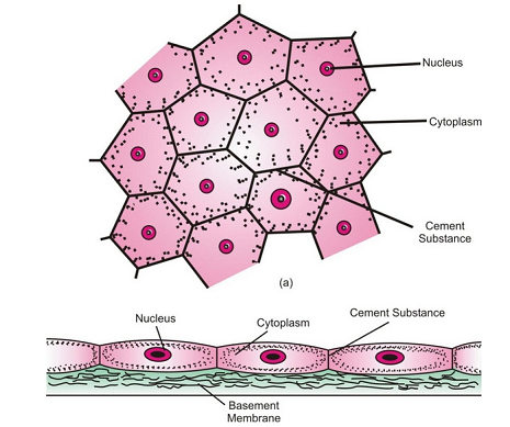
Squamous epithelium (a) Surface view (b) Vertical section.
(b) Cuboidal epithelium
They are cube like cells that fit closely, cells look like squares insection, but free surface appears hexagonal.
It is found in kidney tubules, thyroid vesicles & in glands (salivary glands, sweat glands).
It forms germinal epithelium of gonads (testes & ovaries)
It involves in absorption, excretion & secretion. It also provides mechanical support.

Cuboidal epithelium
Columnar epithelium:
(i) Structure : It consists of single layer of pillar-like cells.
(ii) Occurrence : The columnar epithelium lines the stomach, intestine, gall bladder, etc
.

Columnar epithelium
(d) Ciliated epithelium
- Structure : It consists of cuboidal or columnar cells that develops protoplasmic outgrowth called cilia on their free surfaces.
- Occurrence : Cuboidal ciliated epithelium lines certain parts of urinary tubules of the kidney. Columnar ciliated epithelium lines the nasal passage, oviducts, etc.

Ciliated columnar epithelium
Glandular epithelium:
This epithelium consists of columnar cells modified to secrete chemical sometimes a portion of the epithelial tissue bolds inward and a multicellular gland in formed. It lines the glands such as gastric glands, intestinal glands, etc.

Glandular epithelium
Functions of Epithelial Tissue
- Squamous epithelium (both types) provides protection to underlying parts (organs) against mechanical injury, drying up, entry of germs, etc. It also helps in excretion, gaseous exchange, etc.
- Cuboidal epithelium helps in protection, mechanical support, absorption, excretion, etc.
- Columnar epithelium helps in absorption, secretion and protection. Columnar epithelium of intestine is meant for absorption of water and digested food.
- Ciliated epithelium helps in movement of mucus, urine, eggs, sperms, etc.

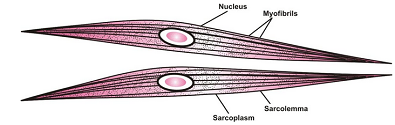
MUSCULAR TISSUE:
Characteristics:
Muscles or muscle tissues consists of long cells. They are also called muscle fibres due to their elongated structure. The muscle cells are arranged parallely and contain special contractile protein which contract and relax in a definite direction. This brings about movement of body parts or limbs and locomotion of organism.
Types of Muscle Tissue
The muscle fibres are classified into three types.
(a) Striated Muscle or Skeletal Muscle: They are also called as voluntary muscles because these are under the control of one’s will. Muscle fibres or cells are multinucleated and unbranched. Each fibra enclosed by thin membrane which is called as sarcolemma.
Cytoplasm is called as sarcoplasm. These Muscles get tired & need rest.

Striated muscles
Non striated muscles: They are involuntary muscles also called as smooth muscles. These muscle fibres are uninucleated & spindle shaped. They are not enclosed by membrane but many fibres are joined together in bundles. Such muscles are found in the walls of stomach, intestine, urinary bladder, bronchi, iris of eye etc. peristaltic movements in alimentary canal are brought about by smooth muscles.
Smooth muscle fibres
Cardiac Muscles:
(i) Structure
- They are so called because they are present only in the wall of the heart. They show striations.
- The fibres are short, cylindrical, branched and joined end to end to form a network.
- Each fibre is surrounded by sarcolemma and has a centrally located nucleus.
- Inter calated discs occur between the ends of fibres.
- Cardiac muscles are involuntary muscles and show rhythmic contraction and relaxation throughout life.
(ii) Occurrence : They are found in the wall of heart only.

Cardiac muscles
Functions of Muscle Tissue:
- Striated muscles help in the movement of the body parts (arms, legs, neck) and locomotion.
- Unstriated muscles help in involuntary activities like passage of food through alimentary canal, flow of air through respiratory tract, flow of blood through the vessels, extrusive movements in urinary bladder, etc.
- Cardiac muscles bring about beating of heart and pumping of blood.

CONNECTIVE TISSUE:
Characteristics:
The name connective tissue suggests that it serves binding and joining of one tissue to another so that there is no interference between activities of different organs. The cells of connective tissue are living and slightly spaced, which in embedded in an intercellular non-living gel-like matrix.
Types of Connective Tissue:
Depending on the type of the matrix, jelly-like, dense (rigid) or fluid connective tissue is further classified.
(a) Connective Tissue Proper (Jelly-like matrix)
It is further divided.
(i) Areolar Tissue
- It is the most widely distributed connective tissue.
- It consists of jelly-like matrix, numerous fibres (white collagen fibres and yellow elastic fibres) and cells.
- This tissue fills spaces inside organs and is also found around blood vessels and nerves, between skin and muscles in bone marrow, etc.

Areolar connective tissue
(ii) Adipose Tissue
- This tissue consists of matrix packed with large, spherical fat cells (adipocytes) filled with fat globules.
- The matrix also contains macrophages, collagen and elastic fibres.
- Adipose tissue is found beneath the skin, in the covering of heart, kidney, etc.
(iii) White Fibrous Tissue
- The matrix has compactly arranged white fibres forming parallel bundles.
- There are few cells lying between the fibres.
- This tissue forms tendons which connect muscles with bones and has great strength but limited flexibility.
|
(iv) Yellow Elastic Tissue
- This tissue has abundant yellow fibres and a few cells in the matrix.
- It form ligaments, which join bones together and has considerable strength and elasticity.

Attachment of tendons and ligaments
|
Attachment of tendons and ligaments |
(b) Skeletal Tissue (Dense or rigid matrix)
It is further classified into two types
(i) Cartilage
- The matrix is composed of proteins and sugars slightly hardened by calcium salts.
- The cells are spaced wide by spaced and get surrounded with fluid filled chambers called lacunae. Cartilage cells are called chondrocytes.
- The surface of cartilage has irregular connective tissue called perichondrium.
- Blood vessels and nerves are absent in the matrix of the cartilage.
- It occurs at the end of long bones, pinnae, end of nose trachea, larynx, etc.

Cartilage
|
Cartilage |
(ii) Bones
- The hard matrix of bone is strengthened by fibres and hardened by calcium and phosphorus salts.
- Bone cells (osteocytes) are contained in lacunae which are arranged in concentric circles.
- The lacunae are traversed by nerves and blood vessels.
- Bones form the endoskeleton of vertebrates.

(a) T.S of long bone; (b) A bone cell
(c) Fluid or Vascular Tissue (fluid or liquid matrix)
It is further classified as
(i) Blood
- It is the most important fluid connective tissue.
- The matrix of this tissue is called plasma which is a straw coloured fluid. The plasma contains 90% water and 10% of various organic and inorganic substances. The organic substances include proteins (albumin, globulin and fibrinogen), carbohydrates, lipids, etc.
- The blood has three types of cells.
- The Erythrocytes or Red blood corpuscles (RBC) are small biconcave cells without nucleus. They are packed with a transport protein called haemoglobin (red in colour) which carry O2 and CO2 to different parts of the body.
- The Leucocytes or White blood corpuscles (WBC) are round or irregular in shape. They possess lobed nucleus. They are colourless and may or may not have granules in their cytoplasm. There are five types of leucocytes-Eosinophil, Basophil, Neutrophil, Lymphocyte and Monocytes.
- The platelets are irregularly shaped, non-nucleated fragments of giant cells.
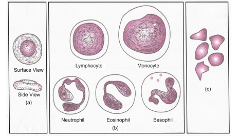
Human blood corpuscles (a) Erythrocytes (RBC); (b) Leucocytes (WBC); (c) Platelets
(ii) Lymph
∙ Lymph is a colourless fluid similar in composition to blood except that it lacks RBC’s, proteins and contains less calcium and phosphorus. In lymph, WBCs are found in abundance.
| The WBC’s exhibit amoeboid movement and the ones present in lymph can reach any part of the body and hence they form the defence system of the body. |
.png)
Functions of connective Tissue:
- Connective Tissue Proper
- Areolar tissue supports internal organs and helps in repair of tissues.
- Adipose tissue stores fat, acts as an insulator.
- White fibrous tissue (tendons) and yellow elastic tissue (ligaments) serve to bind muscles to bones and bone to bone respectively.
- Skeletal Tissue
- Cartilage absorbs stresses, provides flexibility to the body parts and smoothens surface at joints.
- Bones anchors the muscles and provide levers for movement, support the body and for soft body parts and protect many delicate tissues and organs.
- Vascular Tissue
- (i) Blood
- Plasma serves the function of transport (nutrients, hormones, waste products, CO2, etc), regulates water-balance and body temperature.
- RBC helps in transport of O2.
- WBC acts as soldiers and scavengers.
- Platelets help in blood clotting.
- (ii) Lymph protects the body against infection and also transports nutrients.

NERVOUS TISSUE
- They are highly specialized tissue due to which the animals are table to perceive and respond to the stimuli.
- Their functional unit is called as never cell or neuron.
- Cell body is cyton covered by plasma membrane.
- Short, hair like extensions siring from cyton are dendron which are future subdivide into dendrites.
- Axon is long, tail like cylindrical process with fine branches at the end. Axon is covered by a sheath.
- Axon one neuron is very closely placed to the dendrons of another neuron to carry impulses from one to another neuron in the from of electrochemical waves. This close proximity is called as synapse

A neuron
Function of Nervous Tissue
Though all cells possess the ability to respond to change in environment (stimulus), but the Cells of Nervous tissue receive and transmit stimulus very rapidly from sense organs to brain and spinal cord in the form of impulses. This message is interpreted by the brain and spinal cord and the message for response is transmitted to the organs. The functional combination of nerve and muscle tissue enables multicellular animals to move rapidly in response to stimuli.

Comments
Post a Comment